
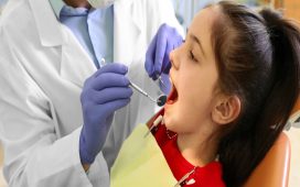
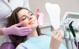
Radiographs, or dental X-rays, play a vital role in modern dentistry, serving as a cornerstone of diagnostic and treatment planning across various specialties, including general, restorative, and cosmetic dentistry. By providing detailed images of teeth, bones, and surrounding tissues, radiographs enable dental professionals to accurately diagnose and treat a wide range of oral health issues, from cavities and gum disease to complex restorative and cosmetic concerns.
This article explores the significance, benefits, and advancements of radiographs in dentistry.
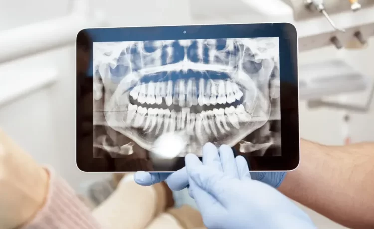
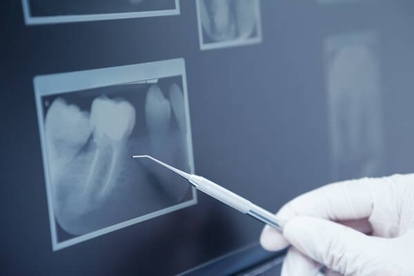
Understanding radiographs
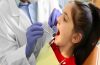
Radiographs, commonly referred to as dental X-rays, are non-invasive diagnostic images that utilize ionizing radiation to produce high-resolution pictures of teeth, bones, and surrounding tissues. These images provide valuable information about the internal structure of the mouth, enabling dental professionals to visualize hidden cavities, cracks, fractures, and other oral health issues.
Types of radiographs
- Intraoral Radiographs: Capture detailed images of teeth and surrounding tissues.
- Periapical radiographs: Show the entire tooth, from crown to root.
- Bitewing radiographs: Reveal interproximal areas between teeth.
- Occlusal radiographs: Display the entire dental arch.
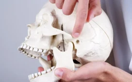
- Extraoral Radiographs: Provide broader views of facial bones and teeth.
- Panoramic radiographs: Show the entire mouth in a single image.
- Cephalometric radiographs: Analyze skull and facial structure.
- Cone Beam Computed Tomography (CBCT): 3D images of teeth, bones, and tissues.
Exploring the importance of radiographs in dentistry
Radiographs provide valuable information about the internal structure of teeth, bones, and surrounding tissues. These include:
Diagnostic Importance
- Early Detection of Cavities: Radiographs help identify cavities in early stages, preventing further decay.
- Hidden Pathologies: Radiographs reveal hidden problems, such as abscesses, cysts, and bone loss.
- Periodontal Disease Diagnosis: Radiographs assess bone level and surrounding tissues.
- Crack and Fracture Detection: Radiographs identify cracks and fractures not visible during visual exams.
Treatment Planning
- Accurate Restorations: Radiographs guide the placement of fillings, crowns, and bridges.
- Dental Implant Planning: Radiographs ensure optimal implant placement.
- Endodontic Treatment: Radiographs visualize root canal anatomy.
- Orthodontic Treatment: Radiographs assess tooth position and growth.
Monitoring Progress
- Treatment Evaluation: Radiographs assess treatment effectiveness.
- Disease Progression: Radiographs monitor disease progression or regression.
- Post-Treatment Follow-Up: Radiographs verify treatment success.
Forensic and Legal Importance
- Identification: Radiographs aid in identifying human remains.
- Bite Mark Analysis: Radiographs help analyze bite marks in forensic cases.
- Legal Documentation: Radiographs provide evidence in dental malpractice cases.
Other Importance
- Patient Education: Radiographs educate patients about their oral health.
- Record Keeping: Radiographs maintain accurate patient records.
- Research and Development: Radiographs contribute to dental research and advancements.
Limitations and risks of radiographs
Although radiographs form the essence of dental diagnosis and treatment, there are some limitations and risks involved:
Limitations
- Image Distortion: Patient movement, incorrect positioning, or equipment malfunctions can distort images.
- Limited View: Radiographs may not capture the entire dental anatomy.
- Overlapping Structures: Radiographs may not distinguish between overlapping teeth or bones.
- Artifacts: Foreign objects (e.g., jewelry, fillings) can interfere with image quality.
- Interpretation Errors: Requires skilled professionals for accurate analysis.
- Limited Sensitivity: May not detect early stages of disease or small lesions.
- Two-Dimensional Representation: Radiographs provide 2D images of 3D structures.
Risks
- Radiation Exposure: Ionizing radiation can increase cancer risk.
- Genetic Damage: Radiation can cause genetic mutations.
- Thyroid Radiation: Exposure to the thyroid gland can increase cancer risk.
- Pregnancy Risks: Radiation exposure during pregnancy can harm fetal development.
- Allergic Reactions: Rarely, patients may be allergic to radiographic materials.
- Infection Risk: Poor infection control can spread diseases.
Future directions of radiographs
To minimize the risks, radiographs have been evolving in the following ways:
- Digital Radiography: Replaces film with digital sensors, enhancing image quality.
- Cone Beam Computed Tomography (CBCT): Provides 3D imaging for complex cases.
- Artificial Intelligence (AI): Assists in image analysis and diagnosis.
- Low-Dose Protocols: Minimize radiation exposure while maintaining image quality.
Radiographs are an indispensable tool in dentistry, providing valuable insights for accurate diagnosis and treatment planning. Understanding the types, benefits, limitations, and future advancements of radiographs enables dental professionals to optimize their use, ultimately enhancing patient care.


